My first confocal image

Originally posted on 17 Nov 2014

It might not be much.
It might not be particularly good.
It might not show anything significant.
But nevertheless… I have run my first confocal image sequence. And I have proof!
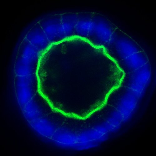
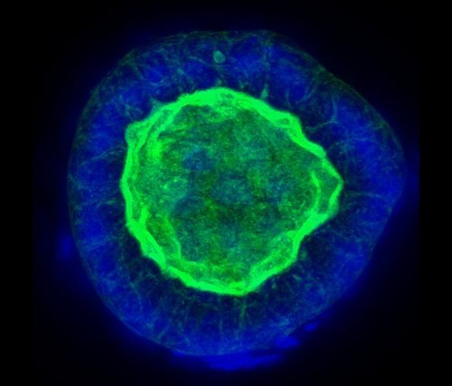
I present to you: a Phalloidin/Hoechst stained MDCK cyst!
(both a single slice as a multiple intensity projection of the z-stack)

MDCK cyst (hollow clump of cells) with nuclei in blue and actin in green
Image taken on a Zeiss 710 Confocal microscope at the University of Dundee.
MDCK cyst (hollow clump of cells) with nuclei in blue and actin in green
Image taken on a Zeiss 710 Confocal microscope at the University of Dundee.

More/nicer to come soon!

Leave a Reply

Your email address will not be published. Required fields are marked *

This site uses Akismet to reduce spam. Learn how your comment data is processed.

%d bloggers like this: