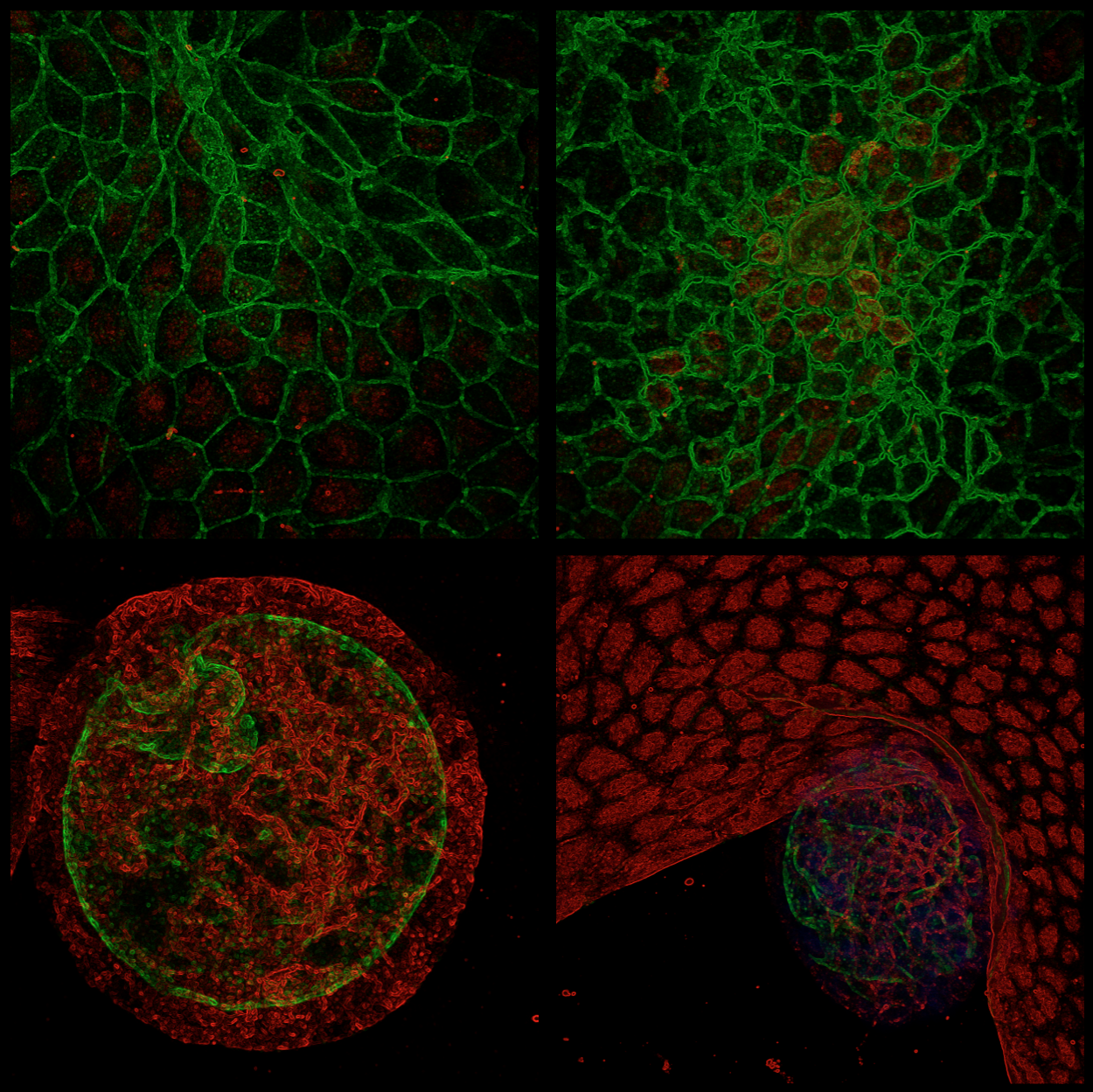
The last images are put though the very professional “Glowing edges” filter found in Microsoft Office Powerpoint.

Science, nerdiness, and hopefully quite some "Eureka!"s
The last images are put though the very professional “Glowing edges” filter found in Microsoft Office Powerpoint.
Hi Val,
Looks really cool. Can you tell a little about what we are seeing and in what context?
Thanks!
(p.s. : Check your mail )
Hi Thomas,
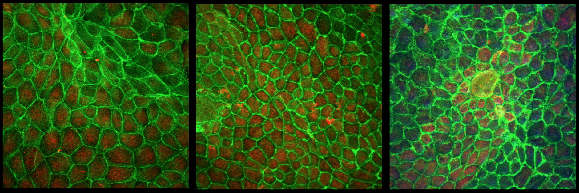
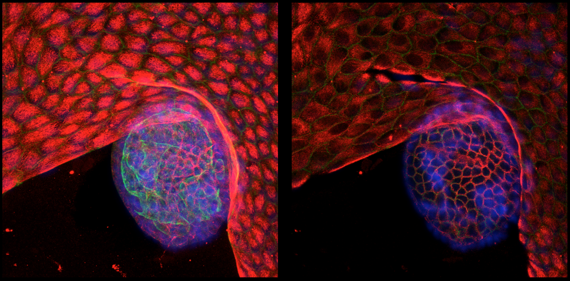
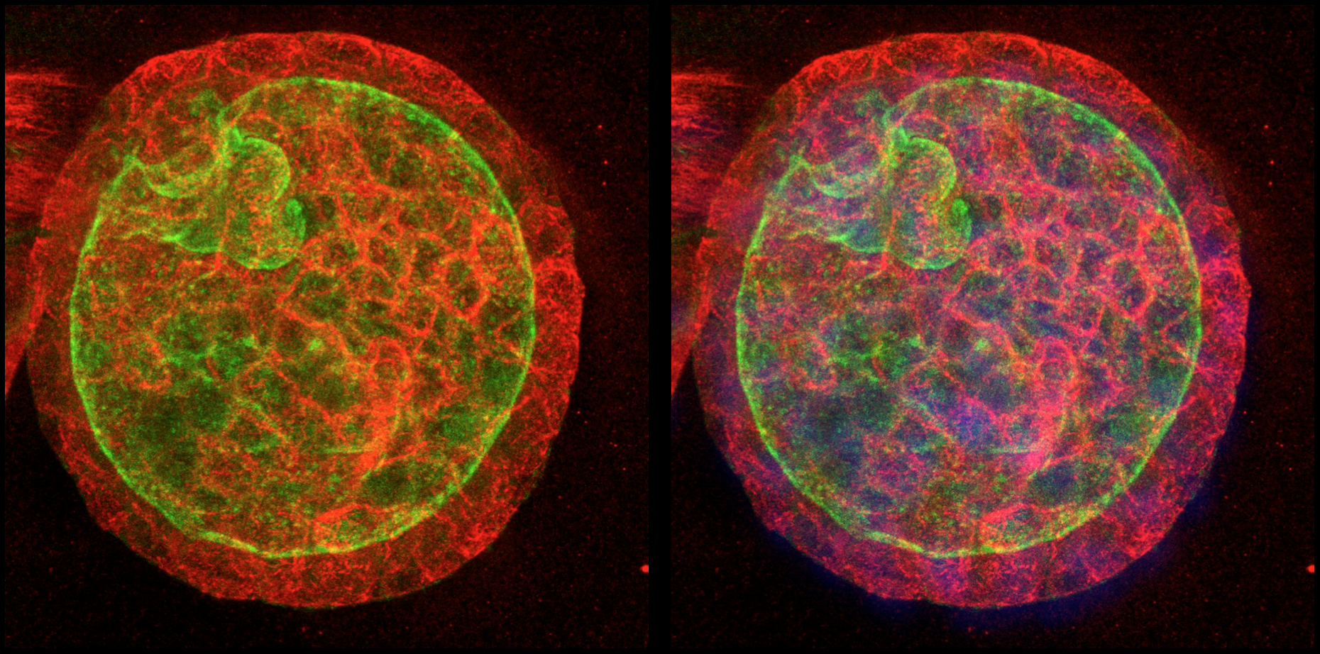
What you see here are MDCK cells (top image) and cysts (middle 2 images). MDCK cells (Madine-Darby Canine Kidney cells) are an immortal cell-line of dog kidney cells. They’re epithelial cells and form round hollow cysts in 3D culture conditions. I’m using them while I establish techniques (because they’re an immortal cell-line, easier to work with, they’re a good model for gut organoids which are my final goal).
I have stained for the nucleus (Hoescht, blue), actin filaments (green) and microtubules (red).
Perhaps this clears things up a bit?